Canine Thymoma: Symptoms, Diagnosis and Treatment
Canine Thymoma: Symptoms, Diagnosis, Treatment, and What Dog Owners Should Know
A canine Thymoma is an uncommon tumor in dogs, but when it occurs, it can cause significant changes in breathing, energy levels, and overall health. Because thymomas grow in the chest cavity—often pressing on the lungs, heart, and major blood vessels—early recognition and veterinary evaluation are essential. Many dogs respond well to treatment, especially when the tumor is detected before it becomes too large.
This comprehensive guide explains what thymomas are, how they affect dogs, the symptoms owners may notice, how veterinarians diagnose them, and the treatment options commonly considered. Understanding this condition can help you make informed decisions and support your dog’s long‑term health.
What a Thymoma Is
A thymoma is a tumor that arises from the epithelial cells of the thymus, an immune‑system organ located in the front part of the chest. The thymus is most active in young animals and becomes smaller with age, but thymic tissue can persist into adulthood. When the cells of this tissue begin to grow abnormally, a thymoma can form.
Thymomas are typically slow‑growing, but their location in the chest means they can cause symptoms even when they are not aggressive. They may compress nearby structures, interfere with breathing, or cause fluid buildup around the lungs.
How Thymomas Affect the Body
Even though thymomas often grow slowly, their position in the cranial mediastinum (the front part of the chest cavity) can lead to several important effects:
Pressure on the lungs, making it harder for the dog to expand the chest fully.
Compression of the heart or major vessels, which may affect circulation.
Fluid accumulation around the lungs (pleural effusion), which can cause sudden breathing difficulty.
Interference with normal immune function, especially if the tumor is associated with autoimmune conditions.
Some thymomas also contain lymphocytes, which can complicate diagnosis and make them appear similar to lymphoma on initial testing.
Symptoms of Thymoma in Dogs
Because thymomas grow inside the chest, many of the symptoms relate to breathing or reduced space for the lungs to expand. Some dogs show subtle signs at first, while others develop sudden respiratory distress.
Common symptoms include:
Labored or rapid breathing
Coughing
Exercise intolerance
Lethargy or weakness
Difficulty swallowing
Regurgitation (especially if the tumor affects the esophagus)
Weight loss
Swelling in the face or front legs (from impaired blood flow)
Collapse during activity
Signs of an emergency
A dog struggling to breathe, breathing with an open mouth, or unable to lie down comfortably should be evaluated by a veterinarian promptly. Respiratory distress can progress quickly and may require immediate supportive care.
Thymoma vs. Lymphoma: Why the Difference Matters
Thymomas and mediastinal lymphoma can look similar on imaging because both involve masses in the chest. However, they differ in important ways:
Thymoma arises from thymic epithelial cells.
Lymphoma arises from lymphocytes and is a systemic cancer.
Distinguishing between the two is important because treatment approaches differ. Thymomas are often treated surgically, while lymphoma is typically managed with chemotherapy.
Veterinarians use a combination of imaging, cytology, and sometimes biopsy to determine which condition is present.
How Veterinarians Diagnose Thymoma
Diagnosing thymoma usually involves several steps, because the tumor is located deep within the chest and may resemble other conditions.
Physical Examination
A veterinarian may detect:
Muffled lung sounds
Abnormal breathing patterns
Signs of fluid in the chest
Weakness or muscle changes
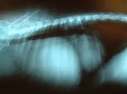
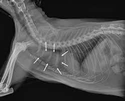
Chest Radiographs (X‑rays)
X‑rays often reveal a mass in the cranial mediastinum. They may also show:
Displacement of the trachea
Fluid around the lungs
Compression of lung lobes
Ultrasound or CT Scan
Advanced imaging helps determine:
The size and shape of the mass
Whether it is invading nearby structures
Whether surgery is feasible
CT scans are especially helpful for surgical planning.
Fine‑Needle Aspirate or Biopsy
A sample of the mass may be collected to examine the cells. However, thymomas can be challenging to diagnose with needle samples alone because they often contain many lymphocytes. In some cases, a biopsy or surgical removal is needed for a definitive diagnosis.
Additional Testing
Depending on the dog’s symptoms, a veterinarian may also evaluate:
Bloodwork
Immune function
Signs of associated conditions such as myasthenia gravis
Thymoma and Myasthenia Gravis
One of the most important associations with thymoma is myasthenia gravis, an autoimmune condition that affects the communication between nerves and muscles. Dogs with myasthenia gravis may experience:
Muscle weakness
Difficulty swallowing
Regurgitation
Megaesophagus (a dilated esophagus that cannot move food properly)
Not all dogs with thymoma develop myasthenia gravis, but the connection is strong enough that veterinarians often test for it when a thymoma is suspected.
Treatment Options for Thymoma in Dogs
Treatment depends on the size of the tumor, whether it has invaded nearby structures, and the dog’s overall health. Because thymomas grow in a confined space, treatment often focuses on removing or reducing the mass to relieve pressure on the lungs and heart.
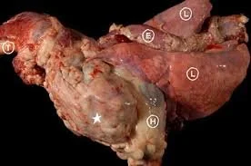
Surgical Removal
Surgery is the most common treatment for thymoma when the tumor is operable. Many thymomas are encapsulated, meaning they have a defined outer layer that makes removal easier. Dogs that undergo successful surgery often experience significant improvement in breathing and quality of life.
Radiation Therapy
If the tumor cannot be fully removed or surgery is not an option, radiation therapy may be considered. Radiation can shrink the tumor or slow its growth, helping relieve symptoms.
Chemotherapy
Chemotherapy is not typically the primary treatment for thymoma, but it may be considered in certain cases, especially if the tumor contains a high number of lymphocytes or if surgery and radiation are not feasible.
Managing Associated Conditions
If a dog has myasthenia gravis or megaesophagus, veterinarians may recommend additional supportive strategies to help manage those conditions.
Prognosis for Dogs With Thymoma
The outlook for dogs with thymoma varies depending on:
Whether the tumor can be surgically removed
Whether it has invaded nearby tissues
Whether complications such as myasthenia gravis are present
The dog’s overall health at the time of diagnosis
Dogs with completely removed thymomas often have a favorable long‑term outlook. Dogs with invasive tumors or severe secondary conditions may have a more guarded prognosis.
Living With a Dog Who Has Thymoma
Caring for a dog with thymoma involves monitoring breathing, watching for changes in energy or appetite, and following the treatment plan recommended by the veterinary team. Many dogs enjoy a good quality of life after treatment, especially when the tumor is detected early.
Owners may find it helpful to:
Track breathing patterns
Monitor for regurgitation or swallowing difficulty
Maintain a calm, low‑stress environment
Schedule regular veterinary check‑ins
Because thymomas can cause sudden changes in breathing, staying alert to early signs of respiratory distress is crucial.
If your dog is showing coughing, breathing changes, or signs of chest discomfort, contacting a veterinarian can help determine what’s causing the symptoms and what care options may support your dog’s health moving forward.